

|
Research projects |


|
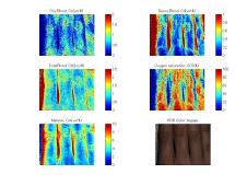
Hemodynamics imaging of human skin tissue by use of digital RGB color photographs |
|
Diffuse reflectance spectroscopy have been widely employed for evaluation of melanin and hemoglobin concentrations in skin tissue that are important for evaluating various pigmented lesions; monitoring tissue metabolism and health status. Multi-spectral imaging technique is a useful tool to extend the diffuse reflectance spectroscopy to the spatial mapping of the chromophores in skin tissue. It can be simply achieved by a monochromatic CCD camera system with narrow band optical filters and an incoherent white light source. Imaging with broadband optical filters such as RGB digital photograph also can provide the spectral images over the visible wavelength range, without the mechanical rotation of filter wheel. |
|
We study a method to visualize the concentrations of melanin, oxygenated blood, and deoxygenated blood distributed in the skin tissue by use of a digital RGB image. In vivo results indicate the ability to acquire the spatiotemporal hemodynamics of subsurface skin tissue. We expect to further extend this work to study the evaluation of vasodilatation or vasoconstriction response to reactive hyperemia such as the endothelial functions of blood vessel. |
|
Click image for loading a movie. |
|
Upper arm was occluded at pressure of 250mmHg by a cuff for 5 min, and then, the cuff was deflated for 5 min. |
|
Mailing address:2-24-16, Naka-cho, Koganei, Tokyo 184-8588 JAPAN TEL:+81-42-388-7065 FAX:+81-42-388-7065 E-mail:inishi@cc.tuat.ac.jp |


|
BASE Bio-Applications and Systems Engineering |
|
BMPL Biomedical photonics Laboratory |

|
TUAT Tokyo University of Agriculture and Technology |
|
Collaboration with Prof. K. Niizeki (Yamagata University) |
|
Copyright © 2007-2013 Medical photonics group at TUAT. All right reserved. Unauthorized use prohibited. |
|
Medical Photonics Group |
|
Biomedical Photonics Laboratory |