

|
Research projects |


|
Visualizing depth and thickness of a local blood region in skin tissue using diffuse reflectance images |
|
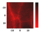
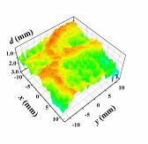
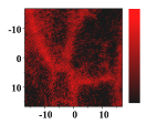
The depth profile of a local blood region in skin tissue is an important parameter in clinical diagnosis and treatments in dermatology. Various vascular malformations have their own histopathological pattern throughout the depths of lesions and are classified by the depth profile of lesions. In laser treatment of port wine stains, the depth profile of a lesion may be used to optimize the pulse duration and radiant exposure of light. Conventional methods such as magnetic resonance imaging and ultrasound imaging have been utilized for differential diagnoses of such diseases, but they often provide insufficient contrast between different types of soft tissue. We proposed a method for visualizing the depth and thickness distribution of a local blood region in skin tissue using diffuse reflectance images at three isosbestic wavelengths of hemoglobin: 420, 585, and 800 nm. Monte Carlo simulation of light transport specifies a relation among optical densities, depth, and thickness of the region under given concentrations of melanin in epidermis and blood in dermis. Experiments with tissue-like agar gel phantoms indicate that a simple circular blood region embedded in scattering media can be visualized with errors of 6% for the depth and 22% for the thickness to the given values. In-vivo measurements on human veins demonstrate that results from the proposed method agree within errors of 30 and 19% for the depth and thickness, respectively, with values obtained from the same veins by the conventional ultrasound technique. Numerical investigation with the Monte |
|
Carlo simulation of light transport in the skin tissue is also performed to discuss effects of deviation in scattering coefficients of skin tissue and absorption coefficients of the local blood region from the typical values of the results. The depth of the local blood region is over- or underestimated as the scattering coefficients of epidermis and dermis decrease or increase, respectively, while the thickness of the region agrees well with the given values below 1.2 mm. Decreases or increases of hematocrit value give over- or underestimation of the thickness, but they have almost no influence on the depth.
|
|
Mailing address:2-24-16, Naka-cho, Koganei, Tokyo 184-8588 JAPAN TEL:+81-42-388-7065 FAX:+81-42-388-7065 E-mail:inishi@cc.tuat.ac.jp |


|
BASE Bio-Applications and Systems Engineering |
|
BMPL Biomedical photonics Laboratory |

|
TUAT Tokyo University of Agriculture and Technology |
|
x (mm) |
|
x (mm) |
|
Depth (mm) |
|
Thickness (mm) |
|
0.5 |
|
1.5 |
|
0 |
|
0.8 |
|
Copyright © 2007-2013 Medical photonics group at TUAT. All right reserved. Unauthorized use prohibited. |
|
Medical Photonics Group |
|
Biomedical Photonics Laboratory |